Diagram Of Hip.and Back.muscles - Muscles Of The Hips And Thighs Human Anatomy And Physiology Lab Bsb 141 : Learn with flashcards, games and more — for free.
Diagram Of Hip.and Back.muscles - Muscles Of The Hips And Thighs Human Anatomy And Physiology Lab Bsb 141 : Learn with flashcards, games and more — for free.. The human back extends from the buttocks to the posterior portion of the neck and shoulders. Nine may seem like quite a lot, but these muscles are essential for creating the wide range of hip movements used by dancers, sportspeople and music lovers. Supraspinatus muscle back workout routine sternocleidomastoid muscle muscle diagram body diagram latissimus dorsi scapula muscular system what are the causes of low back muscle spasming? Now label the diagram in your workbook! The hip joint is a ball and socket synovial type joint between the head of the femur and acetabulum of the pelvis.
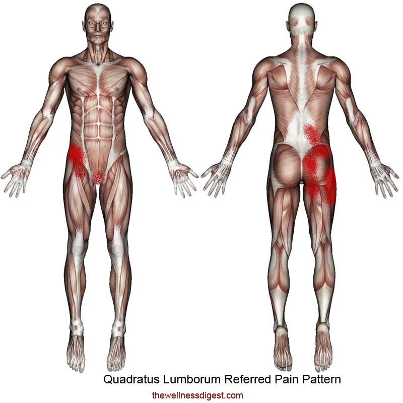
Because this muscle inserts onto the back of the greater trochanter, it produces lateral rotation at the hip. Low back muscle spasming is common because lumbar extensor muscles must contract. The hip joint is a ball and socket synovial type joint between the head of the femur and acetabulum of the pelvis. Some muscles connect to more than one bone or to more than one place on a bone, and therefore have more than one origin. Note that the legs lean backward to keep someone with good posture stands or sits in such as way that their center of gravity lies directly above the pivot point in their hips, thereby avoiding.
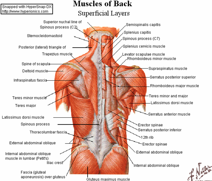
Muscles of the back can be divided into superficial, intermediate, and deep group.since the all the back muscles originate in embryo (fetus) form by locations other than the back, muscles in the.
Human muscle system, the muscles of the human body that work the skeletal system, that are under voluntary control, and that are concerned with movement, posture, and balance. Decreases the angle of a joint; Learn with flashcards, games and more — for free. The veins of the upper portion of the back drain into the. Gluteus maximus, biceps femoris, semitendinosus, semimembranosus at the back and the. Designers also selected these stock illustrations. Muscle tendons in the knee joint and the shoulder joint are crucial in stabilization. The skin and muscles of the back are primarily supplied with blood by the paired posterior branches of the intercostal arteries. Muscles of back of hip an… category: Muscles of the back can be divided into superficial, intermediate, and deep group.since the all the back muscles originate in embryo (fetus) form by locations other than the back, muscles in the. Next to it on both sides of the body is the internal oblique. Back view of muscles, skeleton, organs, nervous system. The muscles of the back can be divided in three main groups according to their anatomical position and function.
It joins the lower limb to the pelvic girdle. Muscles of the posterior … category: Anatomical diagram showing a front view of muscles in the human body. Now label the diagram in your workbook! .lower extremity muscle anatomy lower extremity muscle anatomy, lower extremity muscle anatomy mri, lower extremity muscle anatomy quiz, lower limb muscles anatomy ppt, muscle anatomy of lower extremity, human muscles, lower extremity muscle anatomy, lower extremity muscle.

Most modern anatomists define 17 of these muscles, although some additional muscles may sometimes be considered.
Anatomical diagram showing a front view of muscles in the human body. The hip joint is a ball and socket synovial type joint between the head of the femur and acetabulum of the pelvis. The hip muscles are all the muscles that act on the hip joint. Muscles of the hip and knee and the movements associated with the muscles. Muscles of back of hip an… category: Most modern anatomists define 17 of these muscles, although some additional muscles may sometimes be considered. The human muscular system is complex and has many functions in the body. As we mentioned before the abdominal muscles together participate to maintain your erect posture and prevent hyperlordosis pf vertebral column, hence the william protocol of spine flexion has a positive effect on lumbar hyperhidrosis, back pain, increased flexibility of hip flexor and back extensions. Francesca salvador msc last + show all. The muscles of the back can be divided in three main groups according to their anatomical position and function. The human back extends from the buttocks to the posterior portion of the neck and shoulders. Learn with flashcards, games and more — for free. The core muscles are those in the abdomen, back, and pelvis, and they also stabilize the body and assist in tasks, such as lifting weights.
.lower extremity muscle anatomy lower extremity muscle anatomy, lower extremity muscle anatomy mri, lower extremity muscle anatomy quiz, lower limb muscles anatomy ppt, muscle anatomy of lower extremity, human muscles, lower extremity muscle anatomy, lower extremity muscle. Anatomical diagram showing a front view of muscles in the human body. Several muscles cross the front of the hip and create hip flexion, pulling the thigh and trunk toward each other, but probably the most important is the iliopsoas. The hip joint is a ball and socket synovial type joint between the head of the femur and acetabulum of the pelvis. Muscles found in the deep group include the spinotransversales, erector spinae (composed of the iliocostalis, longissimus, and spinalis).
Muscle tendons in the knee joint and the shoulder joint are crucial in stabilization.
Hip and thigh muscles (overview diagram). The gluteus medius, gluteus minimus, piriformis, tensor fasciae latae on the outside. .lower extremity muscle anatomy lower extremity muscle anatomy, lower extremity muscle anatomy mri, lower extremity muscle anatomy quiz, lower limb muscles anatomy ppt, muscle anatomy of lower extremity, human muscles, lower extremity muscle anatomy, lower extremity muscle. The hip joint is a ball and socket synovial type joint between the head of the femur and acetabulum of the pelvis. Muscles of the back can be divided into superficial, intermediate, and deep group.since the all the back muscles originate in embryo (fetus) form by locations other than the back, muscles in the. Now label the diagram in your workbook! The core muscles are those in the abdomen, back, and pelvis, and they also stabilize the body and assist in tasks, such as lifting weights. The veins of the upper portion of the back drain into the. Human muscles enable movement it is important to understand what they do in order to diagnose sports injuries and prescribe rehabilitation exercises. Everyone should list the structures within muscle. Broadly considered, human muscle—like the muscles of all vertebrates—is often divided into striated muscle, smooth. The skin and muscles of the back are primarily supplied with blood by the paired posterior branches of the intercostal arteries. Note that the legs lean backward to keep someone with good posture stands or sits in such as way that their center of gravity lies directly above the pivot point in their hips, thereby avoiding.


Komentar
Posting Komentar